 | ||||
What Is a
Bacterial Endospore?
Endospores are hardy, defensive structures that enable some bacteria to survive harmful environmental conditions, such as starvation, high temperatures, desiccation (drying out), chemical disinfectants and extremes in pH.
Article Summary: Some bacteria are able to produce tough, dormant structures called endospores which allow them to survive when stressed.
Bacterial Endospores & Vegetative Cells
SPO VIRTUAL CLASSROOMS
 | ||||||
Page last updated:
4/2016
Each endospore is produced by a vegetative cell, an active bacterial cell that undergoes metabolism, divides and goes about the daily business of being alive.
Which Bacteria Can form Endospores?
The ability to form endospores, a process called sporulation, is a rare talent. Bacteria that can do this neat trick are few, but include the notable genera Clostridium and Bacillus. See the Wikipedia article on endospores for a list of endospore-forming bacterial genera.
How are Endospores Formed?
When a vegetative cell of an endospore-forming bacteria detects that essential nutrients are running out it begins to sporulate, a process that takes about 8-10 hours and results in the formation of one endospore.
Steps of sporulation include:
- bacterium's DNA chromosome replicated (is copied)
- cell's plasma membrane pinches off between the replicated chromosomes, forming the forespore
- a second membrane encloses the forespore, with calcium and dipicolinic acid forming a cortex between the inner and outer membrane
- an external spore coat encloses the endospore
- endospore is released once the vegetative cell that generated it dies and disintegrates
See Page 2 for videos on how to Endospore Stain bacteria!
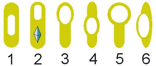
Examples of How Different Types of Bacteria Form Endospores: (1, 4) central endospore; (2, 3, 5) terminal endospore; (6) lateral endospore.
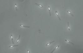
Phase-bright endospores of
Paenibacillus alvei imaged with
phase-contrast microscopy.
John Tyndall (1820 – 1893), prominent physicist, discoverer of endospores and a method used to destroy them, called Tyndallization.
How Were Endospores Discovered?
John Tyndall, a 17th century European physicist, made many contributions to science, including the discovery that some microbes existed in two forms:
- heat-stable form (endospores)
- heat-sensitive form (active, living vegetative cells)
As an endospore, the bacterium is in a dormant, inert state, in which it does not metabolize food (eat) or reproduce, but rather exists in a type of suspended animation, kind of like the seed of a plant.
Tyndallization is still useful for sterilization of growth media in science classes and other situations where modern, expensive autoclaves (instruments that use both heat and pressure to sterilize) are not available for sterilization.
Tyndall found that it took either prolonged or intermittent heating to destroy the resistant heat-stable form. The outcome of his research was a method of sterilizing liquid by heating it to boiling point on successive days, now referred to as Tyndallization.
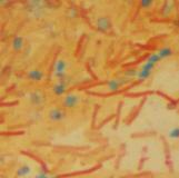
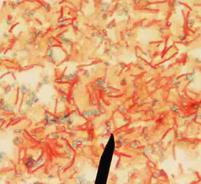
Endospore stained Bacillus subtilis bacteria viewed @ 1000xTM. Vegetative, rod-shaped cells appear red and oval endospores green.
Click here for more
endospore photos.
You have free access to a large collection of materials used in two college-level introductory microbiology courses (8-week & 16-week). The Virtual Microbiology Classroom provides a wide range of free educational resources including PowerPoint Lectures, Study Guides, Review Questions and Practice Test Questions.


