 | ||||
Bacterial Endospores & Vegetative Cells - P2
Staining Bacterial Endospores
Water-based
techniques, such as the Gram stain, will not impart color to endospores. In order to stain these tough structures, the endospore
stain is used.
Sources and Resources
- Bacterial Endospore Formation animated tutorial and quiz from McGraw-Hill.
- How to Do an Endospore Stain Lab Notes article from Science Prof Online Lab Notes article.
- Differential Staining of Bacteria Laboratory Exercise Main Page from the Virtual Microbiology Classroom.
- Endospore Formation animated YouTube video.
- Bacterial Endospores page from Cornell University Department of Microbiology.
- Bacterial Endospores page from Kenyon College's MicrobeWiki.
- Bauman, R. (2012) Microbiology With Diseases by Body System, third edition. Benjamin Cummings.
- Tortora, G., Funke, B. & Case, C. (2013) Microbiology, An Introduction. Pearson.
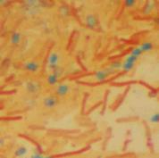
Endospore stained Bacillus subtilis bacteria viewed @ 1000xTM. Vegetative, rod-shaped cells appear red and oval endospores green. Click here for more endospore related photos.
SPO VIRTUAL CLASSROOMS
SPO VIDEO: How to Do an
Endospore Stain
Lab Tutorial
Page last updated: 4/2016
PAGE 2 < Back to Page 1
End of Article
SPO VIDEO: How to Prepare a Bacterial Smear for Endospore Staining
Lab Tutorial
After the primary stain of malachite green is used, the slide is rinsed and the red counterstain safranin is used to impart color to the vegetative bacterial cell. In the end, endospores appear a blue-green color and vegetative cells are red.
During the endospore stain protocol, a bacterial smear of an endospore-producing bacteria, such as Bacillus, is prepared. Then the dye malachite green is forced into the spore with heat from a water bath, in much the same way that fuchsin is forced through the waxy mycolic acid layer of Mycobacterium in the acid-fast stain.
You have free access to a large collection of materials used in two college-level introductory microbiology courses (8-week & 16-week). The Virtual Microbiology Classroom provides a wide range of free educational resources including PowerPoint Lectures, Study Guides, Review Questions and Practice Test Questions.


