 | ||||
How to Prepare & Heat Fix a Bacterial Smear - P2
Staining Bacteria
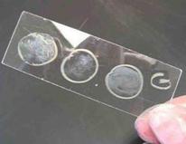
Once a bacterial sample is heat fixed to a microscope slide, there are many staining techniques that can be used to view the bacteria. A simple stain is the use of one dye to increase contrast between the specimen and background when viewed through a microscope.
procedures are more complex, using a series of dyes to distinguish different types of bacteria based on some chemical or structural attribute of the cell.
Differential staining is often used for general identification of bacteria, but does not allow for identification of the exact species.
Page last updated 3/2016
SPO VIRTUAL CLASSROOMS
PAGE 2 < Back to Page 1
Sources & Resources
- Schauer Cynthia (2007) Lab Manual to Microbiology for the Health Sciences, Kalamazoo Valley Community College.
- Bauman, R. (2005) Microbiology. Pearson Benjamin Cummings.
SPO VIDEO
Tutorial on Compound Light Microscope Parts & Operation
When doing a differential stain it is best to use known positive (+) and negative (-) bacterial controls in addition to the unknown bacteria that is being identified. Controls allow for comparison of the unknown to a known bacteria that tests positive for that stain and another that is a known negative.
The following are examples of bacterial controls that could be used in differential stains:
Gram Stain
- + controls: Staphylococcus, Bacillus, Streptococcus
- - controls: Escherichia, Salmonella, Enterobacter
Acid-fast Stain
- + controls: Mycobacteria, Nocardia
- - controls: any type of bacteria that does not have a waxy cell wall
Endospore Stain
- + controls: Bacillus, Clostridium (Clostridium are pathogens, so not a great choice for a control)
- - controls: Any type of bacteria that does not produce endospores
After staining, the bacteria can be viewed using a compound light microscope at a total magnification of 1000xTM.
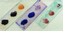
Three slides of stained bacterial smears. Controls are outside circles. Each unknown is in the center. Slides left to right:
SCIENCE VIDEOS
You have free access to a large collection of materials used in two college-level introductory microbiology courses (8-week & 16-week). The Virtual Microbiology Classroom provides a wide range of free educational resources including PowerPoint Lectures, Study Guides, Review Questions and Practice Test Questions.


