 | ||||
Endospore Stain Procedure Lab Notes - P2
Page last updated: 2/2016
Challenges Interpreting the Endospore Stain
If the malachite green stain is allowed to dry out when the slide is on the water bath, a green crust of dye will obscure your specimen. If this has happened, gently tap the crusted dye with your gloved finger while rinsing. This helps dislodge the dried dye.
It should be noted that any debris on the slide can also take up and hold the green stain. Everything that ends up green on the slide is not necessarily an endospore.
Bacterial smear being stained with malachite green over a steaming water bath.
SPO VIRTUAL CLASSROOMS
PAGE 2 < Back to Page 1
Helpful Links on Endospores & Endospore Staining
- Bacterial Endospore Formation animated tutorial and quiz from McGraw-Hill
- What Is a Bacterial Endospore? class notes article from Science Prof Online
- Differential Staining of Bacteria Laboratory Main Page from the Virtual Microbiology Classroom.
- Bacterial Endospore Article from the Department of MIcrobiology, Cornell University.
End of Article
How to Prepare a Bacterial Smear for Endospore Staining
How to Do an Endospore Stain
BACTERIAL SMEAR AND
ENDOSPORE STAIN VIDEOS
You have free access to a large collection of materials used in a college-level introductory microbiology course. The Virtual Microbiology Classroom provides a wide range of free educational resources including PowerPoint Lectures, Study Guides, Review Questions and Practice Test Questions.
Acid-fast Bacteria and the Endospore Stain
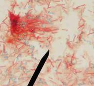
Acid-fast cells, such as members of Mycobacterium and Nocardia have waxy molecules in their cell wall that will take up and retain the malachite green stain when subjected to the endospore staining process.
If the unknown that you have stained has a uniformly green appearance after endospore staining, it may be an acid-fast bacteria. This doesn’t mean that they produce endospores. These are vegetative cells that have taken up color from the heat driving malachite green into their waxy cell wall. With this specimen, a student should also do an acid-fast stain to confirm identification.
Endospores are small, typically oval and you should see numerous uniform examples on your slide. Large or irregular globs of green on the slide may be artifacts.


