 | ||||
Meiosis Classroom Demonstration
with Photos and Assignment
CLASSROOM ACTIVITY from Science Prof Online
eukaryotic cells, particularly the production of gametes, can be a challenging topic for biology students. The following is a step-by-step photographic guide to a simple classroom activity on meiosis that utilizes inexpensive supplies (pipe cleaners, interlocking beads and string).
Article Summary: Here is a classroom hands-on activity to help students practice their understanding of the steps of meiosis. Photo guide and Word doc assignment included.
Meiosis Classroom Activity + Printable Assignment
Supplies Required for Meiosis Activity:
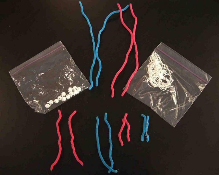
In this exercise, the original cell starts with six chromosomes, so the following supplies are needed.
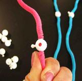
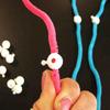
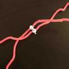
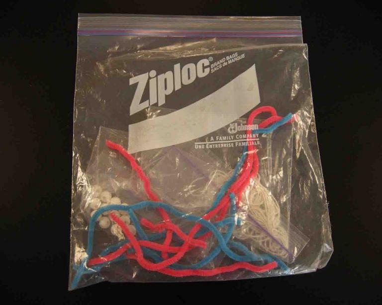
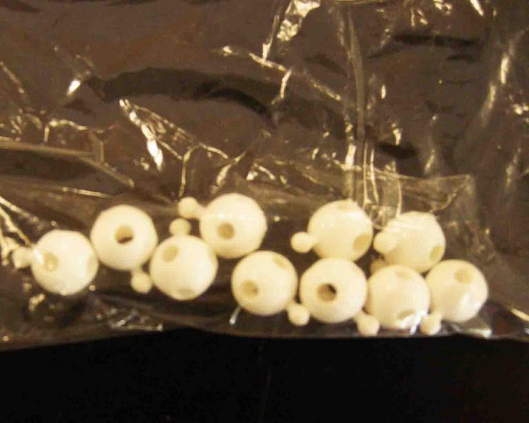
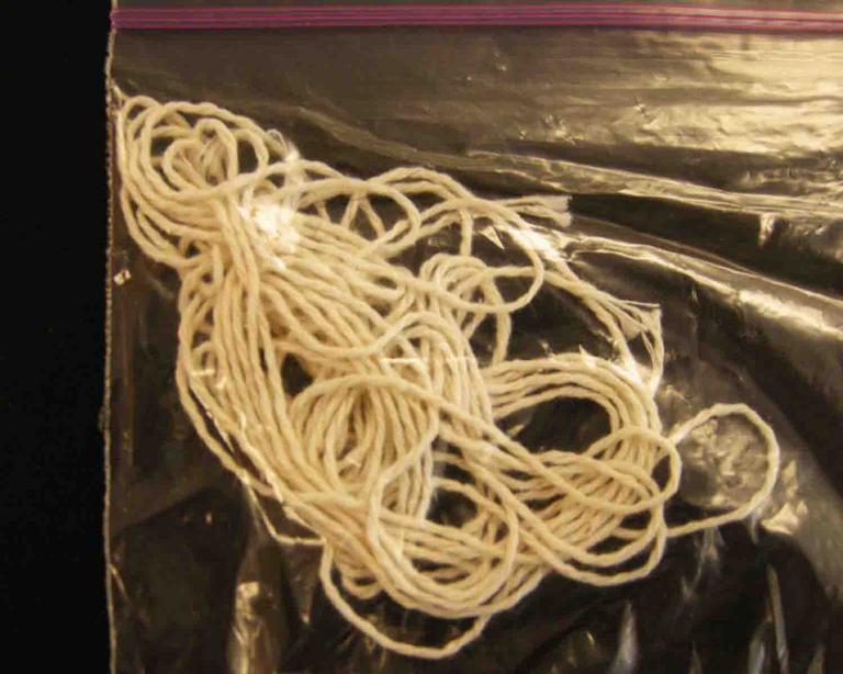
Photos 1 & 2. Large Zip-loc bag contains 12 pipe cleaners (6 blue, 6 pink, of those 4 are long, 4 are medium & 4 are small), interlocking beads (12) and string (2 strands)
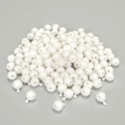
Photos 3 & 4. It helps keep materials organized if interlocking beads and string are each in separate, smaller baggies.
Interphase:
Photo 1. First string one interlocking bead onto each strand of chromatin. Set one of each type of pipe cleaner (6) aside. Those will come into play when DNA replicates.
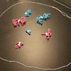
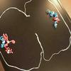
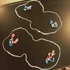
Photo 2. Interphase G1 Phase = six unreplicated, uncondensed strands of chromatin, sting represents the plasma membrane (I don't use additional string to represent the nuclear membrane. It makes exercise too complicated and messy).
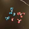
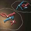
Photo 3. Interphase S Phase = six replicated strands of chromatin (sister chromatids). Snap together the two interlocking beads that represent the centromeres, bringing the other six pipe cleaners into play.
Photo 4. Close-up of replicated chromatin.
CLICK ON PHOTO STRIPS TO SEE ENLARGED PHOTOS.
Meiosis I:
Photo 1. Prophase I, chromatin condenses into chromosomes. Twist pipe cleaners so that they are curly.
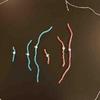
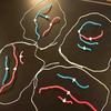
Photo 2. Metaphase I, homologous chromosomes line up at cell equator.
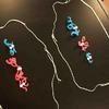
Photo 3. Anaphase I, homologous chromosomes move toward opposite poles of the cell.
Photo 4. Early Telophase I, homologous chomosomes have migrated to opposite end of the cell. Cytokinesis begins.
Photo 5. Late Telophase I after cytokinesis is complete and chromosomes decondense.
Meiosis II:
Prophase II not pictured.
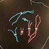
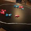
Photo 1. Metaphase II, sister chromatids line up at equator of each cell.
Photo 2. Anaphase II, sister chromatids separate and chromosomes move to either end of the cells.
Photo 3. Early telophase II, chromosomes have migrated to opposite ends of cells, cytokinesis begins.
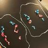
Photo 4. End of meiosis. Four cells with a haploid (n=3)) number of decondensed chromosomes.
More Links to Better Understand Cell Division
- How the Cell Cycle Works, animation & quiz from McGraw-Hill
- Steps of Meiosis, animation & quiz from McGraw-Hill
- How Meiosis Works, animation & quiz from McGraw-Hill
- Comparison of Mitosis & Meiosis, animation & quiz from McGraw-Hill
- Genetics: Tour of the Basics, Genetic Science Learning Center, University of Utah
- Cell Division: Mitosis Lecture Main Page, Virtual Cell Biology Classroom
- Cell Division: Meiosis Lecture Main Page, Virtual Cell Biology Classroom
- Meiosis: Where Sex Starts, video from Crash Course Biology
 | ||||||
SPO VIRTUAL CLASSROOMS
Meiosis Classroom Activity
Page last updated: 1/2016
You have free access to a large collection of materials used in a college-level introductory Cell Biology Course. The Virtual Cell Biology Classroom provides a wide range of free educational resources including Power Point Lectures, Study Guides, Review Questions and Practice Test Questions.
You have FREE access to a large collection of materials used in a college-level introductory biology course. The Virtual Biology Classroom provides a wide range of free educational resources including PowerPoint Lectures, Study Guides, Review Questions & Practice Test Questions.
Where to purchase Interlocking Beads
used in this demonstration:
Carolina Biological Supply: Go to their website (http://www.carolina.com/) and search "DNA beads".
 | ||||
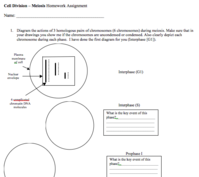
Educators can also download a Word document hand-out of this assignment, called the Meiosis Activity. For more classroom materials on Meiosis, see the Cell Division: Meiosis Lecture Main Page of the Virtual Cell Biology Classroom.
 | ||||
Need a Meiosis Refresher?
